Mammography Screening
Mammography Screening involves using specialized imaging equipment powered by low-dose x rays to check for breast cancer.
Most people are familiar with screening mammograms designed to detect cancer in women of certain ages to identify breast cancer early, even when there are no symptoms.
Mammography also is used to study the breasts of women and men who have signs or symptoms of breast cancer. This typically is called
diagnostic mammography.
HOW MAMMOGRAPHY WORKS
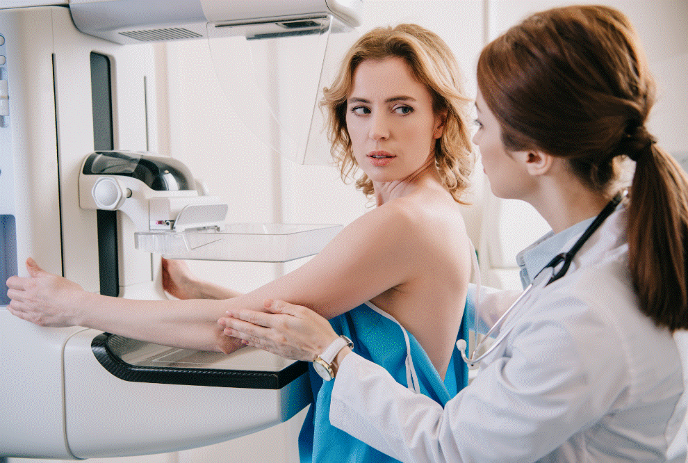
Using equipment designed specifically for mammography, a radiologic technologist position the patient, usually standing, at a special plate used to hold the breast and help capture the image.
Once the patient is in position, the technologist lowers a plastic paddle that compresses or flattens the breast to even its thickness and gets a clear picture of any signs of cancer inside the breast.
Compression also holds the breast to prevent movement that might blur the image and lead to a repeat image.
Most Mammography Screening involves two views of each breast. The technologist releases the paddle quickly and helps to reposition the patient for the next view.
Nearly all mammograms taken today are digital. The image is captured by software in a computer and viewed on a large, high-resolution screen by a radiologist, a doctor who specializes in reading medical images.
The full Mammography Screening examination usually takes only 10 to 20 minutes.
A diagnostic mammogram can take longer but usually focuses on the breast with signs or symptoms or magnifying an area that looks suspicious on a screening mammogram.
According to a standard description on a report that goes to the patient and her physician, the radiologist notes any suspicious findings.
SCREENING RECOMMENDATIONS
Many women affected by breast cancer have no symptoms or signs of disease. Because breast cancer cells can begin spreading throughout the body (called metastasis) soon after a small cancer form, it is important to catch and treat breast cancer early.
A mammogram can show signs of early cancer inside the breast. So, the purpose of screening mammography is to detect early tumors and arrange treatment to prevent cancer spread and death.
As more is learned about breast cancer, mammography, and other breast imaging methods, recommendations about screening mammography have changed, causing some confusion for women and doctors.
As of fall 2018, the American Cancer Society recommended women at average risk for breast cancer:
- consider beginning yearly screening mammograms for women 40 to 44 years old
- have yearly mammograms if 45 to 54 years old
- switch to mammograms every two years once a woman turns 55 or continue yearly mammograms
- continue screening as long as a woman is healthy and expected to live 10 years or more
BENEFITS AND DRAWBACKS
Although Mammography Screening is the most efficient method for detecting early cancer in most women, it is not a perfect tool.
Younger women usually tend to have denser breasts than older ones. This means they have more fibrous tissue.
Some cancers are more difficult to see in mammograms of women with dense breasts.
Some women at higher risk for breast cancer, mostly because of a history of the disease in their close relatives, should have a magnetic resonance imaging (MRI) breast examination along with mammography.
Another potential harm is a high rate of false-positive results from mammograms.
This means the radiologist finds something that looks suspicious for breast cancer and suggests additional imaging or a biopsy, but the results are negative; the woman does not have cancer.
This can cause unnecessary anxiety and testing. False-positive results are more likely to occur in younger women.
Mammograms also expose women to radiation, although the levels are low. There is very low harm from the level received in a single examination unless a woman has repeated mammograms and other x rays.
Compressing the breast tissue means less radiation is needed to get a clear image. Pregnant women should notify the technologist before having a mammogram.
There are proven benefits to Mammography Screening, especially for women 40 years old or older.
The images can show small, early cancers in the breast and the breast’s milk ducts. The test is relatively fast and inexpensive and causes no side effects.
In addition to improved mammograms with digital technology, breast imaging centers also can add special software called computer-aided detection, or CAD, to spot abnormal areas and point them out to the radiologist.
A technique called digital breast tomosynthesis is a form of 3-D mammography that takes multiple images of the breast and reconstructs them using special calculations.
PREPARING FOR A MAMMOGRAM
It is important to tell the primary care physician about any changes or symptoms in the breast before scheduling a mammogram.
It is best to avoid putting any deodorant, powders, or lotions on the breast and under the arms the day of the mammogram because they can cause spots on the image.
The technologist or a staff member at the imaging center should provide a special gown to change into for the examination.
The breasts can be more sensitive at times such as menstruation, so it can help schedule the mammogram for other times of the month.
After a Mammography Screening
Patients should feel no pain or other side effects following a mammogram.
Federal law dictates that patients and their referring physicians receive a timely written report of the examination results.
If a radiologist finds something suspicious, follow-up imaging might be necessary.
A repeat mammogram or other imaging might be used to help guide a biopsy or check and monitor the breast following biopsy or breast cancer treatment.
Resources
Websites
“Mammograms.” National Cancer Institute. December 7, 2016. https://www.cancer.gov/types/breast/mammograms-fact-sheet#q2 (accessed December 6, 2018).
“Mammography.” RadiologyInfo. February 3, 2017. https://www.radiologyinfo.org/en/info.cfm?pg=mammo (accessed December 5, 2018).
“Mammography.” MedlinePlus. October 22, 2018. https://medlineplus.gov/mammography.html (accessed December 5, 2018).
“What Is a Mammogram?” Centers for Disease Control and Prevention. September 11, 2018. https://www.cdc.gov/cancer/breast/basic_info/mammograms.htm (accessed December 5, 2018).
Organizations
American Cancer Society, 250 Williams St. NW, Atlanta, GA, 30303, (800) 227-2345, https://www.cancer.org/.
American College of Radiology, 1891 Preston White Drive, Reston, VA, 20191, (703)648-8900, [email protected], https://www.acr.org.
National Cancer Institute, 9609 Medical Center Drive, Bethesda, MD, 20892-9760, (800) 422-6237, https://www.cancer.gov.


