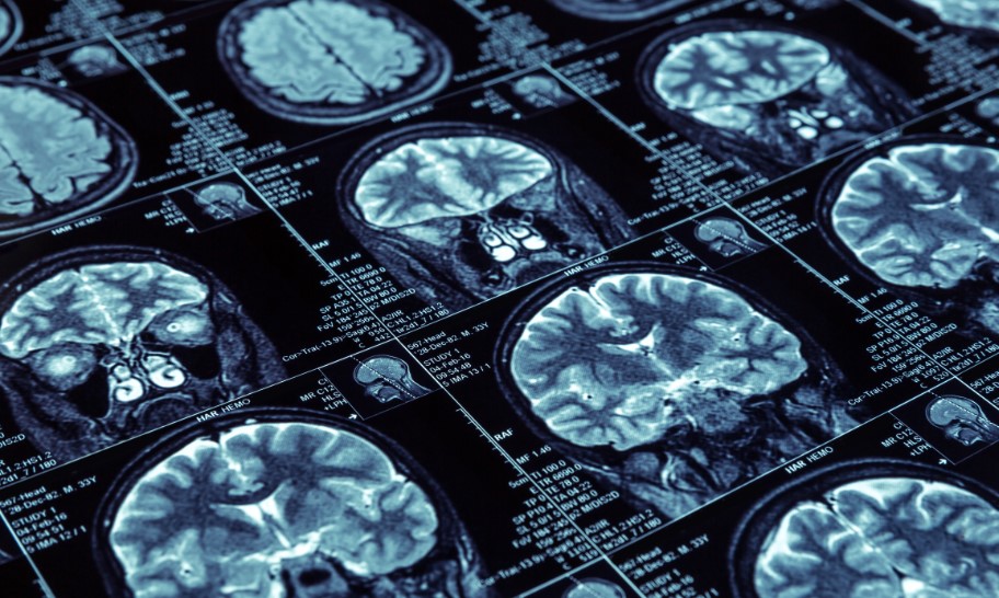
Magnetic resonance imaging (MRI) relates to medical imaging procedures for diagnosing diseases, conditions, or injuries.
MRI uses a strong magnetic field, radio waves, and processing and display by a computer to present detailed images of the body’s tissues, organs, and skeletal system.
MRI scans are advantageous over other imaging techniques because they do not use ionizing radiation, which can damage cells over time. This high-energy type can affect the chemicals in the body’s atoms and lead to cancer in high amounts.
Instead, the energy used to make MRI images comes from a powerful magnet that rotates around the patient and radio waves.
MRI scans are typically performed in radiology departments of hospitals, imaging centers, or even in some physician offices and clinics.
Reasons For Magnetic Resonance Imaging Scans
MRI scanning has many uses, especially for imaging soft tissues in the body.
Examples of common Magnetic resonance imaging uses are knee scans following injury, scans to screen some women at high risk of breast cancer, and detailed images of the heart and blood vessels.
When conducted in a certain way, MRI can also provide images of some of the body’s functions, such as determining how a part of the brain handles functions.
The many forms of applying radio waves and magnets make MRI a helpful way to see inside nearly any body part.
MRI also is used in research, especially of brain function or disease. Examples of uses of MRI scans for patients include:
- checking the brain for signs of stroke
- detection of dementia
- imaging of the spine for illness or infection
- joint evaluation after injuries or for infection and swelling
- evaluate the stomach area for possible tumors or signs of infection
- consider pregnancy to check potential problems with an unborn baby
- breast cancer evaluation in women at high risk for breast cancer
- diseases of the liver
- detect or study tumors in the body
How MRI Exams Work
After undergoing a series of questions and steps to ensure safety in the zone close to the magnet, the patient is asked to lie on a special table attached to the MRI equipment, and a radiologic technologist with special training in MRI moves the table under the magnet using a computer in an adjoining room. The magnet encircles the table or bed.
Intense pulses of radio waves interact with the magnet to temporarily move around protons in the body’s cells.
The protons react differently depending on the type of tissue they make up. Wire coils can be used to send or pick up the electric currents from the patient’s body.
As the machine takes images, the patient will hear loud knocking and clicking sounds. This is normal, and most MRI centers provide headphones to help drown out the equipment’s noise.
The images MRI scans capture show thin cross-sections, similar to slices, which provide excellent detail.
Cautions And Preparation
MRI is a safe examination but can be hazardous if anyone enters the room with a material attracted by the magnet or if the patient has certain kinds of implants in the body.
Patients need to answer all questions on forms or asked by MRI staff to ensure the exam will be safe.
This often includes a thorough description of medical implants such as pacemakers or some dental devices. In general, many metals are affected by magnets.
Also, the machine can interfere with the working of implanted electronic devices. Some materials, even in clothing, can cause burns on a patient.
It is wise to leave valuables such as jewelry at home because patients are escorted when in areas close to the magnet and will be asked to remove jewelry, belts, and other outside devices.
The MRI staff usually provides a safe gown to change into for the exam and see and hear patients in the particular control room’s equipment.
Many MRI exams use a contrast agent injected into the patient’s bloodstream before the examination begins.
The contrast helps highlight blood vessels or areas of the body or brain the physician wants to evaluate.
Child-bearing women and persons allergic to gadolinium contrast should not have contrast during their Magnetic resonance imaging study.
Patients who have severe kidney disease also can experience a severe reaction to the contrast. People with severe claustrophobia might not be able to tolerate a traditional MRI examination.
However, exam times are getting shorter as equipment improves, and there are unique pieces of MRI equipment that are open and less likely to cause claustrophobia.
Likewise, many MRI centers have special goggles or music to help keep children calm and still during their examinations.
MRI Exam Length
MRI scans can vary in length depending on the body part being imaged and the complexity of the examination. However, most MRI exams typically take 30 minutes to an hour to complete. In some cases, particularly for detailed scans or those requiring contrast dye, the exam may take up to 90 minutes.
It’s essential to communicate with your doctor beforehand about how long to expect your specific MRI exam to last.
After An MRI Exam
No special care is needed after a Magnetic resonance imaging exam except for instructions to care for injections or the following sedation is used.
Patients who receive contrast should follow instructions to watch for possible reactions. The part of the body imaged might feel warm but not overly hot.
A radiologist, a doctor, specializing in interpreting MRI and other medical imaging examinations, will review the images and report to the referring physician.
The idea usually is sent to the doctor electronically and viewed on a large monitor. The report should include information on the technique used to capture the images and the findings.
MRI equipment is expensive, and insurance companies might require approval of the examination before it takes place.
Resources
“Information for Patients.” International Society for Magnetic Resonance in Medicine. https://www.ismrm.org/resources/information-for-patients/ (accessed November 28, 2018).
“Magnetic Resonance Imaging (MRI).” National Institute of Biomedical Imaging and Bioengineering. https://www.nibib.nih.gov/science-education/science-topics/magnetic-resonance-imaging-mri (accessed November 28, 2018).
“Magnetic Resonance Imaging.” Radiology Info. https://www.radiologyinfo.org/en/submenu.cfm?pg=mri (accessed November 28, 2018).
American College of Radiology, 1891 Preston White Drive, Reston, VA, 20191, (703)648-8900, info@acr.org, https://www.acr.org.
Society for MR Radiographers and Technologists, 2300 Clayton Road, Suite 620, Concord, CA, 94520, (925) 825-7678, smrt@ismrm.org, https://www.ismrm.org.