DEFINITION
Bladder Ultrasound is a noninvasive test that uses high-frequency sound waves to create an image of the bladder.
PURPOSE
A bladder ultrasound is sometimes called a bladder sonogram. There are a variety of reasons that a bladder ultrasound may be recommended.
Bladder ultrasound can provide images that help a doctor diagnose various diseases and conditions of the bladder, including bladder cancer, urinary tract infections, and kidney stones.
A bladder ultrasound, usually performed with a portable, handheld device, is also often used to evaluate the amount of urine in the bladder before and after voiding.
It can be used to help with bladder rehabilitation by providing feedback about bladder fullness and voiding.
It is also used in nursing homes and extended care settings to help monitor individuals for various bladder diseases and conditions.
PRECAUTIONS
No special precautions are necessary for a bladder ultrasound.
DESCRIPTION
Ultrasound is a non-invasive procedure that is used to provide images of tissue and organs inside the body.
Although the most well-known use of ultrasound technology is to provide images of a fetus inside a pregnant woman, ultrasounds are also used for various other imaging needs in medicine.
They are generally considered preferable to alternative methods when they are feasible and will provide a similar quality picture because they are not invasive and do not involve the use of radiation.
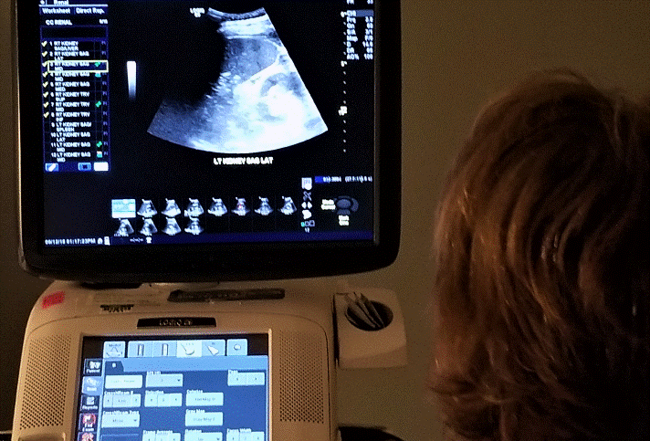
An ultrasound machine generally consists of a computer, recording equipment, and a transducer.
The computer can be a large machine set up permanently in a doctor’s office or clinic, or it can be a handheld device.
Handheld devices are often used in nursing homes and other settings where it is desirable to perform the ultrasound while the individual remains in bed.
The transducer is attached to the computer, and it is the transducer that is moved over the area being imaged by the doctor or technician.
The transducer emits high-frequency sound waves when it comes in contact with the body. High-frequency sound waves are sound waves above the frequency that can be heard by the human ear.
The emitted sound waves bounce off the tissues and organs of the body. They are collected by the transducer, transmitting the information to the computer, where they are translated into visual images and displayed.
Sound waves reflect off of different objects at different rates depending on the density of the object and its distance from the transducer.
It is this information that allows a visual image to be formed. Ultrasound technology allows the images to be displayed in real-time.
In most cases, the images are then recorded and some still frames are also recorded or printed.
When a bladder ultrasound is performed with a traditional ultrasound device, the patient is asked to lie flat on a moveable table that can be adjusted as necessary.
The transducer head is cleaned with alcohol, and a gel is rubbed onto the area to be imaged.
In nursing homes and other care facilities, bladder ultrasounds are generally performed using a handheld ultrasound device while the patient remains in bed.
The gel ensures that the transducer makes good, continuous contact with the skin for best imaging results.
The doctor or technician then slowly moves the transducer over the area of the bladder.
In individuals who are obese or who have excessive skin over the area of the bladder, the skin may need to be moved aside to allow the best imaging of the bladder.
In general, bladder ultrasounds only take a few minutes to perform. After the ultrasound is complete, the gel is wiped off.
In some cases, the results are interpreted as they are displayed. In other cases, images may be recorded and sent to a sonographer or radiologist for interpretation later.
PREPARATION
Loose clothing should be worn on the day of the procedure. In some cases, clothing must be replaced with a gown to allow sufficient access.
Clothing containing metal, jewelry, and any piercing should be removed from the area before the ultrasound.
When the ultrasound is being done to diagnose or monitor a disease or condition, the patient is often asked to drink 32 ounces of water or more before the test.
The patient should finish drinking the water at least one hour before the ultrasound to ensure that the bladder is full and should not urinate until the procedure is complete.
A full bladder helps provide a clear image of the bladder.
AFTERCARE
There is no aftercare required for a bladder ultrasound.
COMPLICATIONS
No complications are expected from a bladder ultrasound. It does not involve radiation of any form, making it preferential to x-rays in many cases.
It also does not have the risk of infection, scarring, or other complications that are often associated with repeated catheterization.
RESULTS
A bladder ultrasound may show a variety of conditions. If the bladder ultrasound was performed to test the ability to void, then the amount of urine remaining in the bladder will indicate whether bladder function is good and void was successful.
Bladder ultrasound may show growths, or other abnormalities that require additional or follow-up diagnostic procedures or that indicate necessary treatment.
HEALTHCARE TEAM ROLES
A nurse or technician generally prepares the patient. A doctor or technician runs the ultrasound machine or uses the handheld device.
In cases where a bladder ultrasound is being performed to monitor or help diagnose a disease or condition, a sonographer or radiologist generally interprets the images.
When the ultrasound is being done to determine bladder fullness or rehabilitation, a nurse may interpret the images.
KEY TERMS
- Transducer— An electronic device that converts sound wave energy into an electrical signal for interpretation.
QUESTIONS TO ASK YOUR DOCTOR
- How much, if any, the fluid should I drink before the ultrasound?
- How long before the ultrasound should I finish drinking it?
- What are you looking for on the ultrasound?
- If the ultrasound reveals a problem, what is the next step?
Resources
“Ultrasound Scan.” Cancer Research UK. May 17, 2019. https://www.cancerresearchuk.org/about-cancer/bladder-cancer/getting-diagnosed/tests-diagnose/ultrasound-scan (accessed June 1, 2020).
“Urinary Tract Imaging.” National Institute of Diabetes and Digestive and Kidney Diseases. April 2020. https://www.niddk.nih.gov/health-information/diagnostic-tests/urinary-tract-imaging (accessed June 1, 2020).
Urology Care Foundation. The Official Foundation of the American Urological Association (AUA). “Ultrasound Imaging.” https://www.urologyhealth.org/urologic-conditions/ultrasound-imaging (accessed June 1, 2020).
American Institute of Ultrasound in Medicine, 14750 Sweitzer Lane, Suite 100, Laurel, MD 20707-5906, (800) 638-5352, http://www.aium.org.
Robert Bockstiegel


