Sensory System
DEFINITION
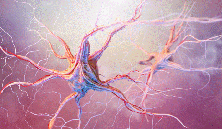
The sensory system is part of the nervous system. It comprises the sensory nerve fibers and receptors of the somatosensory nervous system, the specialized sense organs, and the sensory perception areas of the brain.
The general senses of the somatosensory system include touch, pressure, vibration, proprioception (body position and movement), stretch, temperature, and pain.
The receptors for general senses are nerve endings located throughout the body. The special senses are vision, hearing, equilibrium (balance), and the chemical senses of olfaction (smell) and taste, so-called because they detect chemicals in food and the environment.
Receptors for the special senses are localized and are much more complex than general sense receptors.
Teeth are sometimes classified as special sense organs: nerve endings located in the pulp of teeth are extremely sensitive to stimulation;
however, it is unclear whether teeth have a sensory function other than pain since their sensations of touch are due to nerve endings in ligaments surrounding the teeth.
Different receptor types respond to different types of sensory input or stimuli:
- Electroreceptors detect electrical currents in the environment. This is not found in humans.
- Thermoreceptors respond to temperature.
- Photoreceptors respond to light.
- Mechanoreceptors respond to stretch and sound.
- Chemoreceptors respond primarily to smell and taste.
- Nociceptors are sensory nerve endings that transmit pain sensations. They can be electrical, thermal, mechanical, chemical, or visceral (occurring within the body) receptors.
DESCRIPTION
Anatomy of sensory system
Somatosensory receptors are located in the skin, joints, ligaments, muscles, connective tissues, and organs.
Changes in the environment stimulate exteroceptive receptors in the middle layer of the skin (dermis). Proprioceptive receptors respond to positional changes within the body.
Activation of somatosensory receptors transmits nerve signals through peripheral sensory nerve fibers (axons) to their cell bodies in the dorsal root ganglia of the spinal nerves.
Central processes from the neurons in dorsal root ganglia enter the spinal cord in bundles.
There the bundles split in two: one group transmits proprioceptive impulses through mostly myelinated fibers—axons that are covered with a protective fatty covering called myelin;
the other group contains mostly nonmyelinated fibers that transmit sensations of temperature and pain. Both groups transmit sensations of touch.
The primary somatosensory cortex in the brain integrates this sensory information.
Various types of sensory receptors form the ends of nerve fibers in the skin:
- Hair follicle endings wrap around hair follicles and respond to the displacement of the hair.
- Ruffini endings respond to skin pressure.
- Merkel cells respond to pressure on hairless skin.
- Krause corpuscles respond to pressure on the lips, tongue, and genitals.
- Meissner corpuscles respond to vibrations in the 20-40 hertz (Hz) range in deeper layers of the skin.
- Pacinian corpuscles respond to vibrations in the 150-300 Hz range in deeper layers of the skin.
- Various types of nerve endings throughout the skin respond to temperature, mechanical, and noxious stimuli.
Sensory perceptions are mapped topographically on the cortex of the brain. However, these maps are dynamic and can change when sensory perceptions are lost or gained.
For example, the loss of a finger causes its cortical representation to diminish or disappear and representations of other parts of the hand expand;
cortical representations of a Braille-reading index finger or the fingers of a string musician are expanded compared to their representations in other people’s brains.
The visual sensory system consists of the eyeball, the optic nerve and other sensory nerve pathways, and the visual cortex in the brain that processes visual images.
The eyeball is spherical, about 1 in. (2.5 cm) in length, and consists of three layers.
The external layer of the eyeball consists of the tough, opaque, white sclera and the transparent, dome-shaped cornea. Together, they help protect the eye.
The sclera covers all of the eyeballs except the cornea in front and the optic nerve at the back. The 6 muscles that control eye movement are attached via tendons to the sclera at one end and the eye socket (orbit) of the skull at the other end.
The muscles are controlled by cranial nerves III, IV, and VI. The sclera is covered by a thin mucous membrane called the conjunctiva, which doubles back to form the inner lining of the eyelid.
The eyelids are made up of muscle and connective tissue and are covered with the thinnest skin in the body. The cornea has no blood supply; it absorbs oxygen from the atmosphere through the film of tears.
However, it contains many nerve endings, so damage to the cornea can be excruciating.
The cornea is comprised of five layers:
- The outermost epithelium absorbs oxygen and nutrients and protects against foreign material.
- Bowman’s layer is a transparent sheet of strong protein fibers called collagen.
- The stroma, accounting for 90% of corneal tissue, is 78% water and 16% collagen.
- Descemet’s membrane is a thin, protective collagen layer.
- The extremely thin innermost endothelium pumps excess fluid out of the stroma.
The central layer of the eye consists of the uvea and lens. The uvea consists of the iris, the choroid, and the ciliary body.
The iris is the colored piece of the eye between the cornea and the lens that blocks light. The pupil is the small, dark, round hole in the center of the iris through which light enters the eye.
Circular muscle fibers in the iris contract in bright light to constrict the pupil. Contractile cells in the iris dilate the pupil in the dark. The choroid is a thin membrane that lines the sclera and contains blood vessels for supplying nutrients.
The front of the choroid forms the ciliary body with muscles that act on the iris and on the tiny fibers that form the elastic suspension for the lens called the zonule.
The crystalline lens is the transparent, double-convex structure just behind the pupil. The tiny ciliary muscles of the zonule adjust the shape of the lens to focus on near and far images in a process called accommodation.
The iris divides the space in front of the lens into anterior and posterior chambers. The aqueous humor is the translucent, watery fluid of the anterior chamber continuously produced by the ciliary body and nourishes the cornea and lens.
The aqueous humor is drained into the bloodstream by a system of canals called the trabecular meshwork. Most of the cavity of the eyeball behind the lens is filled with a clear, gel-like vitreous body that exerts constant pressure to maintains the shape of the eye.
The retina—the innermost layer of the eye—is the light-sensitive membrane at the rear of the eyeball that is an outward extension of the central nervous system (CNS).
The sensory or photoreceptor layer of the retina is made up of tightly packed light-sensitive cells called rods and cones, based on their shapes.
The 120 million or so rods contain a single type of light-sensitive pigment. Rods respond to shape, movement, and light and dark.
Rods are most active under low light and are responsible for peripheral and night vision. The six million or so cones respond to bright light and color.
There are three types of cones—red, green, and blue—that are receptors for long, medium, and short wavelengths of light, respectively.
The macula lutea (“yellow spot”) in the middle of the retina, at the exact back of the eye, has the highest concentration of photoreceptor cells—mostly cones.
In the middle of the macula is a tiny pit called the fovea centralis that contains only cones, which are packed tighter than elsewhere in the retina and are responsible for sharp central vision.
Blood vessels and nerve fibers bypass the fovea so that incoming light has a direct path. Each cone in the fovea is directly connected by a single nerve fiber to a single point in the visual cortex, accounting for the clarity of central vision.
Toward the periphery of the retina, the concentration of rods increases, and cones gradually disappear. A single layer of hexagonal cells packed with pigment—the retinal pigmented epithelium (RPE)—lies between the photoreceptor and choroid layers.
The RPE shields the retina from excessive light. The centric retinal artery and vein enter the eyeball through the center of the optic nerve and branch out into arteries, veins, and a capillary network inside the retina.
The optic nerve is created from the axons of photosensitive cells. These axons converge at the optic disk or “blind spot,” where the optic nerve exits the eye.
The outside of the eye is bathed in a three-layer film of tears:
- Meibomian glands produce the outermost oily layer in contact with the air.
- The middle layer, accounting for 90% of the volume of tears, is primarily water, with some salts and proteins. It is produced by the lacrimal gland positioned above the eyeball in the upper outer corner of the orbit.
- The innermost mucin or mucous layer is produced by the goblet cells of the conjunctiva.
Tears flow down and across die cornea into a tiny canal at the inner edge of the eyelid, into the lacrimal sac, and then into the nasolacrimal duct, which opens into the nose.
From there, tears evaporate, drip onto the back of the tongue, where they can be tasted or are reabsorbed by the body.
The ear has three parts: the outer or external ear, the middle ear, and the inner ear or labyrinth.
The pinna or auricle of the outer ear, made of cartilage, is the visible portion. Pinnae are firmly attached to the skull’s temporal bones and move very little so that humans hear better from in front than from behind.
The ear canal or external acoustic meatus is a twisting tube of cartilage and bone covered with skin. Glands within the canal produce cerumen (earwax).
The canal is about 1 in. (2.5 cm) long. It connects the pinna to the tympanic membrane or eardrum—a thin, taut, concave, semitransparent membrane, about 0.33 in. (9 mm) in diameter that separates the outer and middle ear.
The middle ear contains a series of three tiny bones (ossicles): the malleus (“hammer”) is attached on one side to the eardrum and on the other side to the incus (“anvil”); the other end of the incus is attached to the stapes (“stirrup”), the smallest bone in the body.
These three bones mechanically transmit sound vibrations sequentially to the membrane covering the oval or vestibular window opening to the inner ear.
The short eustachian tube connects the middle ear to the throat to equalize air pressure on either side of the eardrum.
The hearing apparatus of the inner ear is the snail-shaped, fluid-filled cochlea. The membrane-covered cochlear or round window is the other opening to the inner ear.
It vibrates in the opposite phase to the vibrations entering through the oval window, moving the fluid in the cochlea.
Inside the cochlea can be found the organ of Corti with about 3,500 susceptible inner hair cells.
Fluid movement within the organ of Corti vibrates the hair cells, which convert the vibrations to electrical signals in the specialized endings of cranial nerve VIII, the vestibulocochlear nerve.
The cochlear branch of the vestibulocochlear nerve extends to the auditory cortex of the temporal lobe of the brain, a place where the nerve impulses are processed and integrated with other nerve impulses to create the sensation of sound.
The brain and ear form a two-way communication system. Nerve bundles transmit instructions from the brain to outer hair cells of the cochlea that calibrate the inner hair cells.
The organ of Corti is encased within the densest bone of the body and floats in the fluid that absorbs accidental vibrations; thus, it is very well protected. In contrast, its hairs are quite vulnerable to damage.
The vestibular or labyrinth portion of the inner ear—consisting of the semicircular canals and otolith organs—is involved in equilibrium.
The bony semicircular canals are arranged at right angles and enclose the fluid-filled, membranous semicircular ducts.
The end of each duct has a bulge called an ampulla with sensory hairs. The otolith organs are hollow sacs in the cavity of the bony labyrinth that is filled with gelatinous endolymph. The inner surfaces of the sacs have hair cells with tiny projections.
Calcium carbonate crystals called otoliths are embedded in the gel over the hair cells.
Olfaction is the sensory response to airborne chemicals breathed into the nose and odors from food and drink that reach the nose through the mouth and pharynx.
Millions of olfactory receptor cells detect odors in the mucous membrane of the roof of each nostril, known as the olfactory epithelium, with a total surface area of about 1 in.2 (5 cm2).
The olfactory epithelium contains Bowman’s glands that produce a complex aqueous secretion that includes odorant-binding proteins. The moist surface of the epithelium dissolves odor molecules from the air to bind to these proteins.
There are 500–1,000 different types of olfactory receptors. A dendrite from each receptor cell body reaches toward the surface of the epithelium and terminates in 8-40 cilia with receptor proteins projecting into the mucus.
Each olfactory neuron is believed to express only one type of receptor. The axons of these sensory neurons terminate in the olfactory bulb.
These olfactory sensory neurons, along with sensory neurons for taste, are the only sensory cells regularly replaced by the body.
Conical mitral cells in the olfactory bulb each synapse with about 1,000 olfactory neuron axons. Mitral cell axons transmit the signals to the region of the forebrain called the rhinencephalon.
Thus, the olfactory bulb is the only neuronal connection from odor reception to perception of smell—a more direct connection than exists for any other sense.
Taste buds are the organs of taste (gustation). They are clusters of 50–150 sensory receptor cells that are often embedded in special structures called papillae projecting from the upper surface and sides of the tongue, the roof of the mouth, and entrance to the pharynx.
There are about 10,000 clusters or taste buds. Women have more than men, and “supertasters” have many more taste buds than other people.
The receptor cells are replaced every one-two weeks. Each receptor in a taste bud responds most strongly to one of the basic tastes.
The tightly packed receptors in each cluster form an opening called a taste pore.
Three different nerves carry taste information to the brainstem: the facial nerve (cranial nerve VII) transports signals from the frontal two-thirds of the tongue;
the glossopharyngeal nerve (cranial nerve IX) carries signals from the hind third of the tongue;
the vagus nerve (cranial nerve X) carries taste information from the epiglottis found at the back of the mouth, although few of these taste fibers persist into adulthood.
From the brainstem, taste signals go to the thalamus and the cerebral cortex. Both olfactory and taste information is also transmitted to the hypothalamus and amygdala of the limbic system.
The trigeminal nerve (cranial nerve V) transmits signals for touch, pressure, temperature, and pain from the tongue to the brain.
SYSTEM SPECIALISTS
Sensory system specialists include:
- ophthalmologists
- optometrists
- ophthalmic nurses, technicians, technologists, and assistants
- opticians
- otolaryngologists
- audiologists
Education, training, and certification of the sensory system
Ophthalmologists are medical doctors (MDs) or doctors of osteopathy (DOs) who are qualified to diagnose and treat eye diseases and perform eye surgeries and other invasive procedures.
As of 2011, there were approximately 200,000 ophthalmologists worldwide, including almost 18,000 in the United States.
After 4 years of college completion, 4 years of medical school, and a licensing exam, they undertake ophthalmology internships and residencies for three-eight years, depending on their area specialization.
A certification given by the American Board of Ophthalmology is granted after passing written and oral exams.
Ophthalmologists practice general ophthalmology or undergo further training, education, and certification in a subspecialty:
- Pediatric ophthalmologists specialize in eye disorders that affect infants, children, and adolescents, including strabismus (crossed eyes), amblyopia (lazy eye), pediatric glaucoma, and genetic disorders.
- Neuroophthalmologists specialize in vision problems related to the nervous system, especially the brain.
- Uveitis and ocular immunologists specialize in benign and cancerous tumors in and around the eye.
- Glaucoma specialists treat increased pressure in the eye—a common disorder among older adults.
- Some ophthalmologists specialize in vitreoretinal diseases, including retinal detachment surgery and vitrectomy (removal of the vitreous).
- Ophthalmic Plastic, cosmetic, and reconstructive surgeons specialize in abnormalities of the eye, eyelid, orbit, and face.
- Ophthalmologists who specialize in cornea and external eye diseases diagnose and treat disorders of the cornea, sclera, conjunctiva, and eyelids and may perform corneal transplants and surgery to correct refractive errors.
- Some ophthalmologists specialize in cataract and/or refractive surgeries.
Optometrists (ODs) are primary eye care doctors who perform physical eye examinations and vision exams.
They diagnose, treat, and manage disorders and diseases of the eye, visual system, and associated structures and diagnose related systemic conditions.
Optometrists complete a bachelor’s degree that includes math and science, followed by four years of optometry school.
Optometry studies include human anatomy and physiology, optics, and optometric sciences, including ocular physiology and pathology, vision anomalies, clinical instruments, general clinical practice, and contact-lens fitting.
Clinical practice under the guidance of teaching optometrists is usually undertaken at school-run clinics in inner-city neighborhoods, nursing homes, or correctional facilities.
Licensed optometrists must pass written and practical examinations administered by state optometry boards. Specialists usually complete an additional one-year internship or residency.
Optometrists sometimes earn masters or doctorate degrees in related medical specialties such as physiological optics, visual sciences, or public health. Practicing optometrists must fulfill state continuing education requirements.
Primary-care or family-practice optometrists treat a wide diversity of patients. They prescribe and often dispense corrective lenses for astigmatism, myopia (near-sightedness), hyperopia (far-sightedness), and presbyopia (age-related farsightedness).
Optometrists also routinely diagnose and treat ocular complications of diabetes and hypertension, prescribe diagnostic and therapeutic medications, and perform certain surgical procedures, including laser refractive surgeries.
They collaborate with ophthalmologists in preoperative and postoperative treatments.
Many optometrists specialize:
- Pediatric optometrists treat common childhood vision problems such as myopia, amblyopia, and strabismus.
- Geriatric optometrists specialize in diagnosing and treating eye disorders of the elderly, such as cataracts, macular degeneration, glaucoma, and diabetic eye disorders.
- Contact-lens specialists perform complex fittings and treat irritations and infections from contact lenses.
- Low vision/vision rehabilitation optometrists work with patients who use specialized equipment, such as magnifiers, charts, telescopes, colored filters, lenses, prisms, computerized devices, lights, and closed-circuit televisions to maximize vision. They provide rehabilitation services for patients with visual complications from brain injury or stroke.
- Vision therapists or developmental vision specialists treat children with amblyopia and strabismus and learning disorders, and behavioral problems related to vision.
Because of their roles in vision screening programs in schools, daycare centers, and other institutions, nurses are often front-line workers in the initial detection of visual problems.
Ophthalmic nurses, technicians, and assistants take patient histories and perform preliminary tests.
Nurses and assistants perform patient education, explain glaucoma medication regimens, teach patients to insert eye drops and help them select lens and frame materials, measure the eyes, and assist with eyeglass fittings.
They assist in testing for macular degeneration and in screening patients for laser eye surgery.
Since many ophthalmologists now operate their own ambulatory surgery centers, nurses and assistants prepare the operating room, prep patients, insert anesthetic eye drops, and assist with surgeries.
Nurses and technicians also instill eye-dilating drops and inject fluorescein dye used in angiography to examine blood vessels.
An ophthalmic technician or retinal photographer takes photographs used for analyzing retinopathies.
Only registered nurses and physicians can prepare the drugs used for photodynamic therapy (PDT).
Because of advances in ophthalmic and optometric technologies, many aspects of eye exams and treatment that were previously performed only by ophthalmologists and optometrists are now performed by allied professionals. Many ophthalmic nurses, technicians, and assistants:
- measure visual acuity (clarity) and refraction under low and high illumination
- examine eye structures using biomicroscopy with pupillary dilation
- determine ocular motility and alignment, binocularity, and visual fields
- measure near the point of convergence—the point as which an object approaching the nose is seen as double—and near fusionai vergence amplitudes
- determine relative accommodation and accommodative amplitude and facility
- examine pupillary responses
- measure depth perception
- measure intraocular pressure (IOP)
In addition to completing the requirements to become a registered nurse, an ophthalmic registered nurse must have at least two years of hill-time experience (4,000 hours) in ophthalmic registered nursing practices before taking a certification examination through the National Certifying Board for Ophthalmic Registered Nurses.
Allied ophthalmic professionals may seek certification through the American Board of Opticianry/National Contact Lens Examiners (ABO/NCLE) or other organizations.
They can progress through certification levels with additional education and work experience and by passing examinations. Certified ophthalmic assistant is the lowest certification level, requiring six months to one year of training.
Certified ophthalmic technicians have, on average, one-two years of training. Certified ophthalmic technologists have more than two years of training. Allied professionals can complete specialized training in certified programs for performing eye exams.
Vision rehabilitation teachers, social workers, psychotherapists, experts in assistive technologies, and others are often involved in low-vision rehabilitation.
Dietitians work with eye disease patients suffering from diabetes, high blood pressure, or heart disease.
Opticians fit, dispense and verify corrective lenses, including eyeglasses and frames, contact lenses, and other optical devices.
Manufacturing opticians or ophthalmic laboratory technicians grind, cut, and edge lenses based on the prescription and frame size and insert lenses into frames.
Dispensing opticians fill corrective lens prescriptions and assist customers in choosing and fitting suitable frames, including measuring the distances between the pupils’ centers and between the eye surface and lens and adjusting the frame to the face.
Opticians also measure the size and shape of eyes for contacts, select appropriate lenses, ensure their proper fit, and instruct clients on their wear and maintenance.
In some states, opticians who fit contact lenses must work under the immediate supervision of an OD or MD.
Specialized opticians, called ocularists, create artificial eyes and shells for patients who have lost an eye.
Some opticians specialize in nonprescription optics such as binoculars or microscopes.
Most opticians earn a two-year associate’s degree from a community college or university. Some opticians are trained through an apprenticeship with a licensed optician or on-the-job training in a clinic or optical shop.
In most states, opticians must pass the National Opticianry Competency Examination of the ABO/NCLE and an additional exam to dispense contact lenses.
The ABO/NCLE also offers advanced certification for ophthalmic dispensing of spectacles, contact lens, and refraction services.
Certification must be renewed every three years. The ABO offers a thesis-requiring master’s degree program for opticians with advanced certification.
Otolaryngologists or ear/nose/throat (ENT) specialists are MDs trained in the medical and surgical treatment and management of disorders and diseases of the ear, nose, throat, and related structures of the neck and head, including the sinuses, larynx, and mouth.
They treat ear infections, ear noise (tinnitus), hearing loss, balance disorders, and certain cranial nerve diseases.
Otolaryngologists also handle congenital disorders of the outer and inner ear, olfactory disorders, and disorders of the head and neck that may affect vision and hearing.
Otolaryngologists complete four years of college and four years of medical school, followed by at least five years of specialized training.
They have to pass an examination to be certified by the American Board of Otolaryngology. Some otolaryngologists pursue a one-or two-year training fellowship in one of eight subspecialties of sensory system:
- allergies
- plastic and reconstructive facial surgery
- head and neck, including benign and cancerous tumors
- otology/neurotology, which includes diseases, injuries, infections, benign tumors, and nerve pathway disorders that affect hearing and balance, as well as tinnitus, swimmer’s ear, hearing loss, dizziness, and ear, face, and neck pain
- pediatric otolaryngology, including congenital disabilities, developmental delays, ear infections, and allergy and sinus diseases
- rhinology or disorders of the nose, including olfactory disorders
- laryngology (throat disorders)
- sleep disorders
Audiologists diagnose and treat disorders of the auditory and vestibular systems and provide preventive and rehabilitative services, including auditory training, speech reading, and listening skills.
They prescribe and fit assistive devices such as hearing aids and telecommunication systems, provide ear-and hearing-related surgical monitoring, and design and implement hearing conservation programs and newborn screenings.
Some audiologists sell and service hearing aids, although most states require that hearing aids be fitted and sold only by licensed specialists such as dispensing audiologists.
Audiologists also research the prevention, assessment, and management of hearing and balance disorders.
Audiologists have at least a master’s degree from an accredited university or professional school of audiology. Most states require audiologists to be licensed.
After completing a nine-month postgraduate clinical fellowship and passing a national standardized examination, an audiologist can earn a Certificate of Clinical Competency in Audiology from the American SpeechLanguage-Hearing Association.
Post-baccalaureate four-year Doctor of Audiology (AuD) programs were restituted in the early 1990s to replace the master’s as the entry-level degree and ress the need for increased clinical training and expanded consumer services.
AuD programs include a 12-month supervised clinical fellowship. However, some audiologists have other doctoral degrees with special training in hearing disorders.
Primary care physicians and other healthcare professionals, as well as audiologists, perform hearing examinations. Speech/language specialists also provide treatment and rehabilitation for the deaf and hard of hearing.
Disclaimer: The pieces of information found in this article are not a tool for self-diagnosis or a substitute for professional care.


